Coomassie-Brillantblau (häufig nur als Coomassie bezeichnet) sind Triphenylmethanfarbstoffe aus der Gruppe der Säurefarbstoffe, die in der Biochemie zum Anfärben von Proteinen verwendet werden. Coomassie-Brillant-Blau ist der am häufigsten verwendete Proteinfarbstoff.[1] Sie führen zu einer sequenzunspezifischen Proteinfärbung. Ursprünglich wurden diese Farbstoffe in der Textilindustrie für Wolle genutzt.
Der Zusatz „R“ im Namen Coomassie Brillant Blau R-250 (C.I. Acid Blue 83) ist eine Abkürzung der englischen Bezeichnung „reddish“, zu deutsch etwa „rötlich“. Der Zusatz „G“ im Namen Coomassie Brillant Blau G-250 (C.I. Acid Blue 90) steht als Abkürzung für „greenish“, zu deutsch also für grünlich. Die Zahl 250 im Namen bezieht sich auf die Farbstärke, mit der der Hersteller Imperial Chemical Industries das Färbevermögen quantitativ beschrieb. Oft wird in Veröffentlichungen nur Coomassie ohne genauere Angabe verwendet. Coomassie bezeichnet nach dem Colour Index über 40 verschiedene Substanzen. Es gibt sogar ein weiteres Coomassie-Blau (Coomassie Blue RL, synonym Acid Blue 92, C.I. 13390), das nach dem Merck Index (10. Auflage) eine völlig andere Struktur besitzt.
| Coomassie-Brillantblau | ||||
| Name | Coomassie-Brillantblau R-250 | Coomassie-Brillantblau G-250 | ||
| Andere Namen | C.I. Acid Blue 83 Brillantblau R C.I. 42660 Xylenbrillantcyanin |
C.I. Acid Blue 90 Brillantblau G C.I. 42655 | ||
| Strukturformel | 
|

| ||
| CAS-Nummer | 6104-59-2 | 6104-58-1 | ||
| PubChem | 61365 | 6324599 | ||
| Summenformel | C45H44N3NaO7S2 | C47H48N3NaO7S2 | ||
| Molare Masse | 825,97 g·mol−1 | 854,02 g·mol−1 | ||
| Aggregatzustand | fest | |||
| GHS- Kennzeichnung [2][3] |
|
| ||
| H- und P-Sätze | keine H-Sätze | keine H-Sätze | ||
| keine EUH-Sätze | keine EUH-Sätze | |||
| keine P-Sätze | keine P-Sätze | |||
Eigenschaften
BearbeitenDie Farbe von Coomassie-Brillant-Blau richtet sich nach dem pH-Wert.[4] Bei pH <0 ist G-250 rot mit einem Absorptionsmaximum bei einer Wellenlänge von 465 nm.[4] Bei pH 1 ist es grün mit einem Absorptionsmaximum bei 620 nm.[4] Bei pH >2 ist es blau mit einem Absorptionsmaximum bei 595 nm.[4] Bei pH 7 ist G-250 blau und hat einen Extinktionskoeffizienten von 43,000 M−1 cm−1.[4] Bei der roten Form (pH <0) sind alle drei Stickstoffatome protoniert und die zwei Sulfonsäuregruppen deprotoniert (Nettoladung +1). Bei der grünen Form ist die Nettoladung 0. Bei pH 7 in der blauen Form ist nur das Stickstoffatom der Diphenylamingruppe protoniert und die Nettoladung −1. Der pKa für die Deprotonierungen liegen bei 1,15 und 1,82.[4] Erst bei sehr hohen pH-Werten wird das letzte Stickstoffatom deprotoniert und die Farbe wird rosa (pKa 12,4).[4] Coomassie-Brillant-Blau bindet nichtkovalent über elektrostatische Wechselwirkungen an Aminogruppen und Carboxygruppen von Proteinen. Die Bindung an Proteine stabilisiert die blaue Form mit der Nettoladung −1 auch bei niedrigen pH-Werten, bei denen die gelöste Form bereits in der roten Form mit der Nettoladung +1 vorliegt.[4] Die Bindung an das Tensid SDS erzeugt einen stabilen Komplex und stabilisiert die grüne Form mit der Nettoladung 0, weshalb SDS eine Störsubstanz bei photometrischen Bestimmungen nach Bradford ist.[5]
-
 Coomassie-Brillant-Blau G-250 in Pulverform
 -
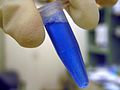 Coomassie-Lösung
 -

-
 Coomassie-gefärbte Proteine nach einer SDS-PAGE
 -
 Coomassie-Brillant-Blau G-250 in Isopropanol


Anwendungen
BearbeitenTypischerweise wird Coomassie-Brillantblau R-250 z. B. zum Einfärben von aufgetrennten Proteinen in einem Polyacrylamid-Gel benutzt,[6] z. B. nach einer SDS-PAGE oder einer BN-PAGE.[7][8] Es wird außerdem in der 2D-Gelelektrophorese zur Sichtbarmachung der getrennten Proteinpunkte verwendet und beim Western Blot zur Angleichung der eingesetzten Proteinmengen.[9] Die Nachweisgrenze für Coomassie-Brillant-Blau R-250 liegt bei ungefähr 0,1 µg Protein pro Bande in einem Gel, für Coomassie-Brillant-Blau G-250 bei etwa 0,5 µg. Mit Coomassie-Brillant-Blau G-250 lassen sich Proteine jedoch deutlich schneller anfärben. Besondere Formulierungen als kolloidale Lösungen verbessern die Nachweisgrenze von Coomassie-Brillant-Blau G-250 auf circa 4 ng Protein pro Bande.[10][11] Coomassie-Brillantblau G-250 dient auch der Quantifizierung des Proteingehaltes wässriger Lösungen nach der Methode von Bradford.[12]
Neben Methylenblau wurde und wird Coomassie-Brillantblau auch zum dauerhaften Einfärben von nicht zum menschlichen Verzehr geeignetem Fleisch und Schlachtabfällen verwendet. Coomassie-Brillant-Blau G-250 wird auch bei Augenoperationen eingesetzt.[13] Aufgrund der reduzierten Toxizität im Vergleich zu bisher verwendeten Farbstoffen (Indocyaningrün oder Trypanblau) werden Triphenylmethanfarbstoffe wie Coomassie-Brillant-Blau G-250, Patentblau V oder C.I. Acid Violet 17 als Vitalfärbung für das Anfärben von natürlich vorhandenen oder krankhaft veränderten Strukturen (Gliose) im lebenden Auge eingesetzt. Mit Hilfe dieser Farbstoffe lassen sich vor allem die transparenten Strukturen der Netzhaut, Hornhaut oder der Linsenkapsel gezielt anfärben und bei Bedarf chirurgisch entfernen. Für die Technik des Anfärbens von Gewebe an der Netzhaut hat sich der Fachbegriff der Chromovitrektomie etabliert.[14][15][16][17][18] Ebenso wird es in der Forensik zur Färbung von Fingerabdrücken verwendet.[19]
Geschichte
BearbeitenDer Name des ursprünglich als Wollfarbstoff entwickelten Coomassie-Brillant-Blau stammt von der afrikanischen Stadt Kumasi in Ghana. Er wurde zur Erinnerung an die britische Besetzung der damaligen Aschanti-Hauptstadt Coomassie – heute Kumasi – im Jahr 1896 gewählt. Der Name bezeichnete eine Gruppe saurer Wollfarbstoffe.[20] Coomassie war ursprünglich eine registrierte Marke von Levinstein Ltd, die ab 1926 von der Imperial Chemical Industries übernommen wurde.[21] Blaue disulfonierte Triphenylmethanfarbstoffe wurden erstmals 1913 von Max Weiler in Elberfeld hergestellt.[22] In den folgenden Jahren wurden verschiedene Synthesen patentiert.[23][24][25] Im Jahr 1963 wurde Coomassie-Brillant-Blau R-250 von Stephen Fazekas de St. Groth zur Proteinfärbung in Elektrophoresen eingesetzt,[26] wo es neben der Silberfärbung zu einer der häufigsten Färbungen von Proteinen in Elektrophoresegelen wurde.[27]
Weblinks
BearbeitenEinzelnachweise
Bearbeiten- ↑ C. Arndt, S. Koristka, A. Feldmann, R. Bergmann, M. Bachmann: Coomassie Brilliant Blue Staining of Polyacrylamide Gels. In: Methods in molecular biology. Band 1853, 2018, S. 27–30, doi:10.1007/978-1-4939-8745-0_4, PMID 30097926.
- ↑ Datenblatt Coomassie-Brillant-Blau bei Merck, abgerufen am 25. Mai 2021.
- ↑ Datenblatt Coomassie-Brillant-Blau bei Merck, abgerufen am 25. Mai 2021.
- ↑ a b c d e f g h H. J. Chial, H. B. Thompson, A. G. Splittgerber: A spectral study of the charge forms of Coomassie Blue G. In: Analytical Biochemistry. 209. Jahrgang, Nr. 2, 1993, S. 258–266, doi:10.1006/abio.1993.1117, PMID 7682385.
- ↑ S. J. Compton, C. G. Jones: Mechanism of dye response and interference in the Bradford protein assay. In: Analytical biochemistry. Band 151, Nummer 2, Dezember 1985, S. 369–374, doi:10.1016/0003-2697(85)90190-3, PMID 4096375.
- ↑ Wilson CM: Studies and critique of Amido Black 10B, Coomassie Blue R, and Fast Green FCF as stains for proteins after polyacrylamide gel electrophoresis. In: Anal Biochem. 96. Jahrgang, Nr. 2, 1979, S. 263-78, PMID 89822.
- ↑ Hermann Schägger, Gebhard von Jagow: Blue native electrophoresis for isolation of membrane protein complexes in enzymatically active form. In: Analytical Biochemistry. 1991, Band 199, Nummer 2, S. 223–231 doi:10.1016/0003-2697(91)90094-a.
- ↑ Ilka Wittig, Hans-Peter Braun, Hermann Schägger: Blue native PAGE. In: Nature Protocols. 2006, Band 1, Nummer 1, S. 418–428 doi:10.1038/nprot.2006.62.
- ↑ C. Welinder, L. Ekblad: Coomassie staining as loading control in Western blot analysis. In: Journal of Proteome Research. Band 10, Nummer 3, März 2011, S. 1416–1419, doi:10.1021/pr1011476, PMID 21186791.
- ↑ N. Dyballa, S. Metzger: Fast and sensitive colloidal coomassie G-250 staining for proteins in polyacrylamide gels. In: Journal of visualized experiments : JoVE. Nummer 30, August 2009, S. , doi:10.3791/1431, PMID 19684561, PMC 3149902 (freier Volltext).
- ↑ D. Kang et al. (2002): Highly Sensitive and Fast Protein Detection with Coomassie Brilliant Blue in Sodium Dodecyl Sulfate-Polyacrylamide Gel Electrophoresis. In: Bull. Korean Chem. Soc. Bd. 23, Nr. 11, S. 1511–1512. doi:10.5012/bkcs.2002.23.11.1511.
- ↑ N. Noaman, J. R. Coorssen: Coomassie does it (better): A Robin Hood approach to total protein quantification. In: Analytical biochemistry. Band 556, September 2018, S. 53–56, doi:10.1016/j.ab.2018.05.012, PMID 29763592.
- ↑ H. Enaida, T. Hisatomi, S. Nakao, Y. Ikeda, S. Yoshida, T. Ishibashi: Chromovitrectomy and vital dyes. In: Developments in ophthalmology. Band 54, 2014, S. 120–125, doi:10.1159/000360457, PMID 25196760 (Review).
- ↑ S. Balaiya, V. S. Brar, R. K. Murthy, K. V. Chalam: Comparative in vitro safety analysis of dyes for chromovitrectomy: indocyanine green, brilliant blue green, bromophenol blue, and infracyanine green. In: Retina 31(6):1128-36 (2011). PMID 21394068.
- ↑ E. B. Rodrigues, M. Maia, C. H. Meyer, E. M. Penha, E. Dib, M. E. Farah: Vital dyes for chromovitrectomy. In: Curr Opin Ophthalmol. 18(3):179-87 (2007). PMID 17435423.
- ↑ A. Iriyama, K. Kadonosono, Y. Tamaki, Y. Yanagi: Effect of Brilliant Blue G on the retinal ganglion cells of rats. In: Retina 32(3):613-6 (2012). PMID 22392093.
- ↑ Y. S. Chang, S. Y. Tseng, S. H. Tseng, Y. T. Chen, J. H. Hsiao: Comparison of dyes for cataract surgery. Part 1: cytotoxicity to corneal endothelial cells in a rabbit model. In: Journal of Cataract and Refractive Surgery. 31(4):792-8 (2005). PMID 15899458.
- ↑ S. Thaler, J. Hofmann, K. U. Bartz-Schmidt, F. Schuettauf, C. Haritoglou, E. Yoeruek: Methyl blue and aniline blue versus patent blue and trypan blue as vital dyes in cataract surgery: capsule staining properties and cytotoxicity to human cultured corneal endothelial cells. In: Journal of Cataract and Refractive Surgery. 37(6):1147-53 (2011). PMID 21596258.
- ↑ E. Brunelle, A. M. Le, C. Huynh, K. Wingfield, L. Halámková, J. Agudelo, J. Halámek: Coomassie Brilliant Blue G-250 Dye: An Application for Forensic Fingerprint Analysis. In: Analytical chemistry. Band 89, Nummer 7, April 2017, S. 4314–4319, doi:10.1021/acs.analchem.7b00510, PMID 28293949.
- ↑ M. R. Fox: Dye-makers of Great Britain 1856-1976 : A History of Chemists, Companies, Products and Changes. Imperial Chemical Industries, Manchester 1987, S. 38.
- ↑ M. R. Fox: Dye-makers of Great Britain 1856-1976 : A History of Chemists, Companies, Products and Changes. Imperial Chemical Industries, Manchester 1987, S. 259.
- ↑ Colour Index. 3rd Auflage. Band 4. Society of Dyers and Colourists, Bradford 1971, S. 4397–4398 (colour-index.org ( des vom 19. Juli 2011 im Internet Archive) [abgerufen am 20. Juli 2010]).
- ↑ FR patent 474260, "Procédé de production de colorants de la série du triarylméthane", erteilt 1915-02-16 an die Bayer AG.
- ↑ US patent 1218232, Weiler, Max, "Blue Triphenylmethane Dye", erteilt 1917-03-06
- ↑ GB patent 275609, "Manufacture of Triarylmethane-dyestuffs", erteilt 1927-11-03 an die IG Farbenindustrie.
- ↑ Stephen Fazekas de St Groth, Robert G. Webster, A. Datyner: Two new staining procedures for quantitative estimation of proteins on electrophoretic strips. In: Biochimica et Biophysica Acta. Band 71, Mai 1963, S. 377–391, doi:10.1016/0006-3002(63)91092-8, PMID 18421828.
- ↑ J. L. Brunelle, R. Green: Coomassie blue staining. In: Methods in enzymology. Band 541, 2014, S. 161–167, doi:10.1016/B978-0-12-420119-4.00013-6, PMID 24674070.